X Ray Record of Spinal Cord
For most people X-rays are safe. Long-term effects after x-ray exposure of the spinal cord.

Cervical Myelopathy Johns Hopkins Medicine
Article in Russian Shirochina OA Morozova TD Vyborov MP.

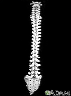
. Asked Apr 4 2017 in Health Biomechanics by Jamaican. Often a full length x-ray including each section of the spine cervical thoracic lumbar sacrumpelvis is needed to understand the global picture. Inflammation of the gray matter of the nerves.
Radiology department of the Alrijne Hospital in Leiderdorp the Netherlands. An x-ray record of the spinal cord using a contrast medium is aan. Inflammation of the gray matter of the spinal cord.
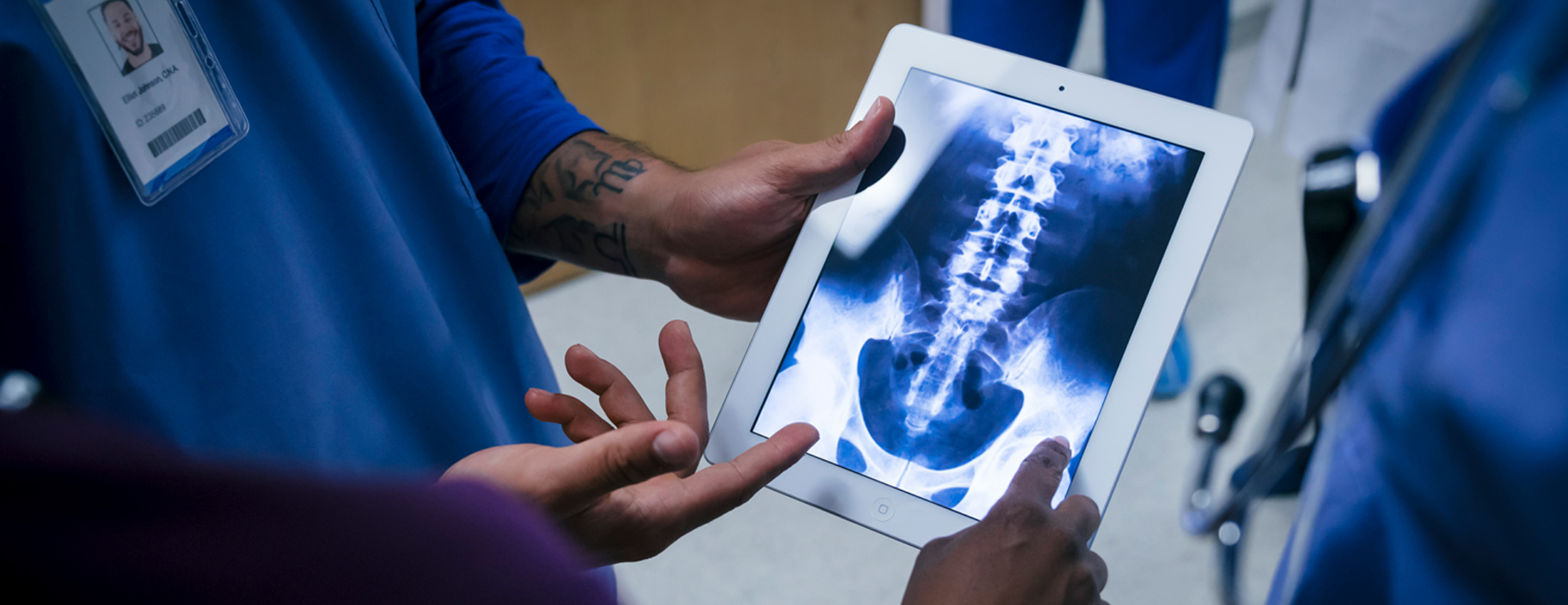
X-ray record image of the spinal cord. Looking at abnormalities of the spinal cord vertebrae. Pertaining to the cerebellum pons.
Rather than treating one focused area or body part we treat the entire person as a whole. Van der Kogel A J. Pulse generator has a battery which is rechargeable via external wireless power charger.
Some worry that the radiation can cause changes in cells that may lead to cancer. X-ray record of the spinal cord. Partner with the Right2Know March and support the common right to know our food sources.
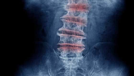
Diagnosing Spinal Cord Injury X-Rays Any evidence of either anterior or posterior displacement 1 between vertebrae greater than 4mm on a lateral cervical radiograph is considered abnormal. Coloured myelogram spinal cord X- ray of a healthy human spinal cord white in the lower lumbar back. X-ray record of the spinal cord In this test Radiopague die or air is inserted in Sub-Arachnoid space then x-rays are taken.
It is lined by spinal pia mater and contained by the other spinal meninges in the thecal sac. It is worth noting though that if you are in any doubt regarding your interpretation of these often. Gomez Lopez J 1.
Even in adults a normal cross-table lateral x-ray does not exclude a spinal cord injury. Spinal cord Nerve Roots Assessing for Back Pain. The radiologist will also use a CT scan when doing a myelogram.
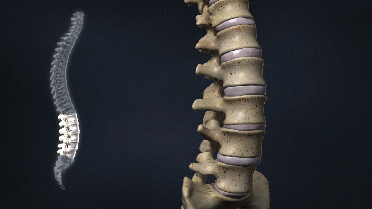
The spinal cord is the part of the central nervous system that is found within the spinal canal of the vertebral columnThe cord extends from the corticomedullary junction at the foramen magnum of the skull down to the tip of the conus medullaris within the lumbar cistern. 3 rows x-ray record of the spinal cord made visible with a radiopaque contrast medium. Disease of the nerves.
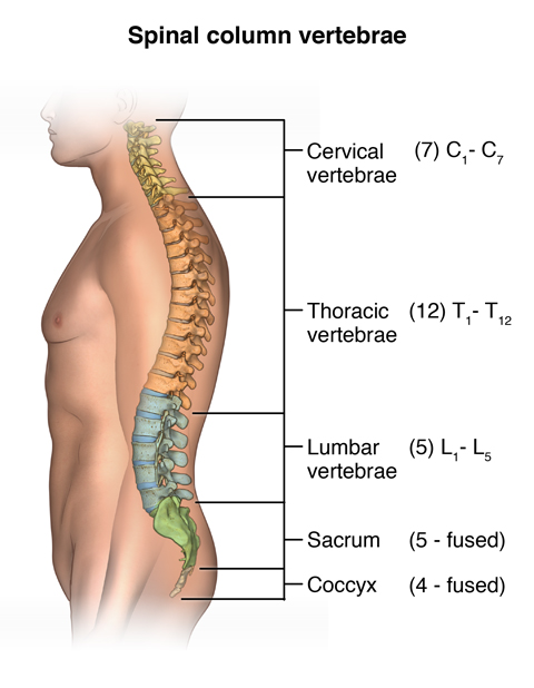
The vertebrae spinal bones orange protect and support the spinal cord. This is an updated version largely based on the recommendations of the combined task forces of the North American Spine Society the American Society of Spine Radiology and the American Society. X ray record of the spinal cord pass answer.
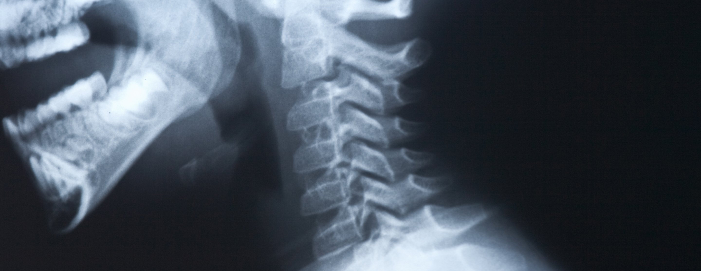
Answered Apr 4 2017 by. Chattanoogas only daily newspaper. Cervical spine X-rays arent something youll see commonly outside of an Emergency Department but the importance of being able to read them and the risks of missing significant findings spinal cord injury death make brushing up on the basics well worthwhile.
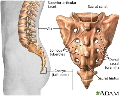
Disease of a spinal nerve root. At the base of the vertebral column is the sacrum the bone that links the vertebrae to the pelvis. Tenderness of the neck and careful neurologic examination must stay the main way of diagnosing a patient especially in the pediatric population.
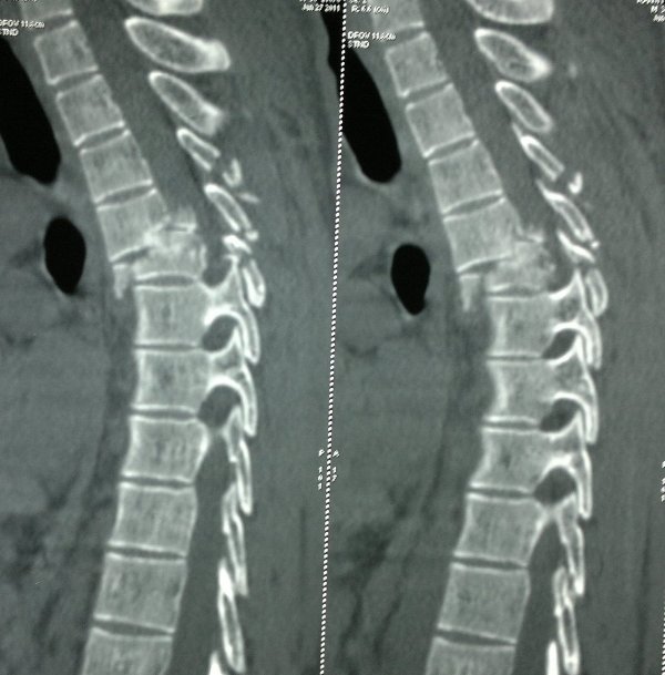
Medical imaging experts have reviewed the patient records of 302 men and women who had a much-needed X-ray of the blood vessels near the spinal cord and found that the procedure often feared for. It is seen from the side left and front right. X ray record image of the spinal cord a.
Subcutaneous pulse generators are a component of the spinal cord stimulators placed in various places of the body. Radiologists should be familiar with the different locations of spinal cord stimulators implantable pulse generators. Anterior displacement of less than half the diameter of the vertebral body suggests unilateral facet dislocation displacement greater than this indicates bilateral facet.
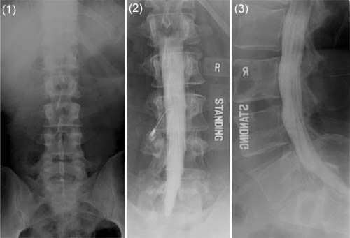
For patients with spinal deformities standing laying and bending x-rays help determine the flexibility. X-ray diagnosis of spinal cord tumors. The contrast dye is injected into the spinal column before the procedure.
But the amount used in spinal X. Complete coverage of breaking news in Chattanooga Other local regional and national news. Spinal x ray webmd.
X ray record of the spinal cord A Electroencephalogram B Electromyogram C from MRMT 1305 at Lone Star College System. Plain radiographs are negative in 25 of pediatric patients with an injury to the spinal cord. The contrast dye appears on an X-ray screen allowing the radiologist to see the spinal cord subarachnoid space and other nearby structures more clearly than with standard X-rays of the spine.
Lumbar Disc Nomenclature 20.
Lumbosacral Spine X Ray

Pin On Justice Clearinghouse
X Rays Of The Spine Neck Or Back Johns Hopkins Medicine
Spinal Cord Injury Presentation And Treatment Bone And Spine
Lumbosacral Spine X Ray Information Mount Sinai New York
Myelography Myelogram

X Ray Stock Photos Royalty Free Images Vectors Video Spinal Cord Injury Cell Therapy X Ray
X Rays Of The Spine Neck Or Back Johns Hopkins Medicine

Radiological Imaging Of Lung And Spinal Cord A Chest X Ray Pa Erect Download Scientific Diagram

Disc Osteophyte Complexes Opll And Compressive Myelopathy Due To Canal Stensosis Spine Surgery Radiology Osteophyte

X Rays Of The Spine Neck Or Back Johns Hopkins Medicine
Lumbosacral Spine X Ray Information Mount Sinai New York
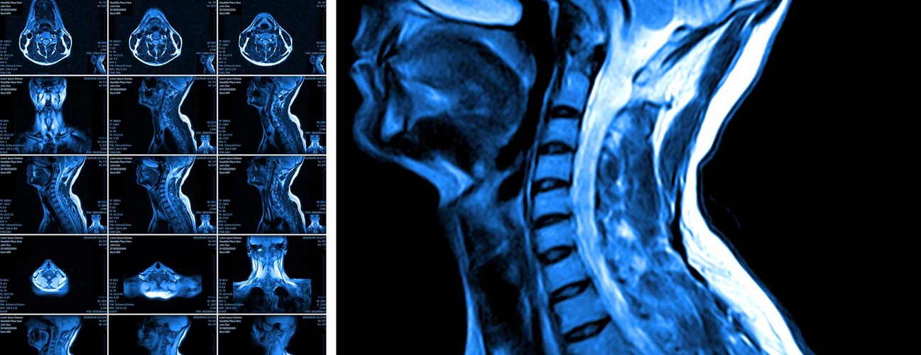
Cervical Myelopathy Johns Hopkins Medicine
Neck X Ray

Thoracic Spine X Ray

Pin By Koester Bradley Llp On Koester Bradley Llp Infographics Neck Injury Traumatic Brain Injury Spinal Cord Injury
Pin On Latest News
Lumbar Spinal Stenosis Johns Hopkins Medicine

Assesment Of The X Ray Features Of Lumbar Disc Degeneration Lateral Download Scientific Diagram


Comments
Post a Comment